Leukemia cells
SECTIONS
I. Cancer
– Cancer profile of 9/11
– Statistics of first responders
– Types of cancer in nuclear events
(- Body parts most vulnerable to radiation)
(- Leukemia)
(- Malignant solid tumors)
– Does jet fuel cause cancer?
– Similarities with Hiroshima and Nagasaki
– Dose of radiation and symptoms
– Fallout and cancer risk
– Elevated fission products at the WTC site
– Elevated tritium at the WTC site
– Asbestos and Silverstein’s white elephant
– Clean up efforts at the WTC
– Shielding
– Protection for workers
– Depleted uranium and cancer risks
– Afghanistan
– Chernobyl
– Analysis of the WTC dust
– WTC’s toxic clouds
– Black rain
II. Other (non-cancer) health effects
(- Types of radiation and damage they do)
(- Acute radiation injuries)
(- Chronic radiation illnesses)
– Human effects of atomic explosion
– Blast effects – overpressure
– Blast pressure and negative pressure
– Flashburns
– Glass injuries
– Damage from the heat ray
– Keloids
– Cataracts
(- Body parts most vulnerable to radiation)
(- Leukemia)
(- Malignant solid tumors)
_________________________________________________________________
INTRODUCTION
There is no denying the high incidence of cancer among the first responders at the World Trade Center. In Jun 16, 2006, almost five years after the September 11 attacks, it was reported that 283 WTC rescue and recovery workers have been diagnosed with cancer. Thirty-three have died from cancer. Even though there has been a virtual media blackout on this issue, the prevalence of cancer among the first responders has reached alarming proportions. The cancer profile of the first responders mimics that of people exposed to radiation. There are many similarities between the 9/11 cancer-sufferers and the cancer-sufferers of Hiroshima and Nagasaki. There are also startling similarities with the cancer victims of nuclear power station accidents. WTC workers are being struck with brain cancers, lymphomas, leukemias and breast cancers. There are some unusual cancers appearing with abnormally high frequency as well, such as Non-Hodgkin’s lymphoma. Other workers who do not have cancer suffer from illnesses that stem from damage to their immune system. Out of 7,300 Ground Zero workers involved in a lawsuit, 41 have died as of 2006. Some of the deaths are due to brain cancer, a cancer associated with nuclear radiation exposure. Exposure to the dust cloud that formed in the aftermath of the destruction of the World Trade Center seems to be a risk factor in the development of serious illnesses of Ground Zero workers. These sick and dying people are living proof that nuclear devices were detonated on 9/11. Their cancers are a red flag that there is more to 9/11 than the official story. The epidemic of cancer is an important piece of the puzzle that is 9/11. By fitting this piece into the puzzle, a more complete picture of 9/11 emerges. And it is a picture that shows a nuclear detonation took place at the World Trade Center on 9/11.

IMAGE: Radiation symbol [From Answers.com: Radiation Poisoning]
CANCER PROFILE OF WTC WORKERS
Like the cancer victims of Hiroshima and Nagasaki, blood cell cancers take the center stage in the cancer profile of the WTC workers. WTC workers are being struck down with leukemias, lymphomas and myelomas. They are suffering from rare and unusual cancers in higher frequency than the normal population. They are also developing solid tumors such as brain cancer, lung cancer, breast cancer, thyroid cancer and doing so in alarmingly high numbers.
“To date, 75 recovery workers on or around what is now known as “the Pile”—the rubble that remained after the World Trade Center towers collapsed on the morning of September 11, 2001—have been diagnosed with blood cell cancers that a half-dozen top doctors and epidemiologists have confirmed as having been likely caused by that exposure.” [Village Voice – Death by Dust]
Leukemia
Acute white blood-cell cancers are rare in males under age 40 in normal circumstances. However when people are exposed to radioactive fallout such as in Hiroshima and Nagasaki, these blood cell cancers are seen more often in this age group.
Leukemia is a cancer that is called the sentinel-disease for radiation exposure as it is one the first cancers to appear in such groups. The difference in rates between the irradiated population and the non-irradiated population is large.
“One in 150,000 white males under 40 would normally get the type of acute white blood-cell cancer” [Fire Chief – Cancers being found in 9/11 responders 6/16/2006]
After 9/11, we have 75 workers out of a population of 50,000 rescue and cleanup workers with this type of cancer (as of 2006). This is equal to 245 people out of 150,000 getting acute white blood cell cancer. Compare this to the figure of 1 person out of 150,000 getting acute white blood cell cancer in the white male under-40 group. The increase in acute white blood-cell cancers in the WTC worker cohort is of the order of 24,400%, or to put it another way, the incidence of acute white blood cell cancer in the WTC rescue and cleanup population is greater than the incidence of acute white blood cell cancer occuring in the normal white male population by a factor of 240. The spike in acute white blood cell cancers among the WTC workers indicates that something unusual happened at the WTC site during the 9/11 attack that caused the skyrocketing of these cancers.
Leukemia flared up in both Hiroshima and Nagasaki 3 years after the blasts, hitting a peak around 1951. By 1978, the rate of death from leukemia proved to be about twice as high had the atomic bomb not been dropped. (Bochkov and Oftedal, 330). [The Medical Implications of Nuclear War. Washington, DC: National Academy Press, 1986.]
The following tables show the excess deaths from radiation-induced leukemia and from radiation-induced cancers of all types among a long-term cohort of Hiroshima and
| Cause of death | Total number of deaths | Estimated number of deaths due to radiation | Percentage of deaths attributable to radiation |
|---|---|---|---|
| Leukemia | 176 | 89 | 51% |
| Other types of cancer* | 4,687 | 339 | 7% |
| Total | 4,863 | 428 | 9 |
*Solid cancers, such as stomach, lung, breast, colorectal and liver cancers
| Leukemia | Other cancers* | ||||
|---|---|---|---|---|---|
| Distance from hypocenter (km) | No. of persons | No. of deaths | Percent attributable to radiation | No. of deaths | Percent attributable to radiation |
| <1 | 810 | 22 | 100% | 128 | 42% |
| 1.0-1.5 | 10,590 | 79 | 64% | 1156 | 18% |
| 1.5-2.0 | 17,370 | 36 | 29% | 1622 | 4% |
| 2.0-2.5 | 21,343 | 39 | 4% | 1781 | 0.5% |
*Solid cancers, such as stomach, lung, breast, colorectal and liver cancers
What Is Leukemia?
Leukemia comes from a Greek word meaning “white blood” and is often referred to as cancer of the blood. The term refers to a group of closely related malignant conditions affecting the immature blood-forming cells in the bone marrow.so is that certain cells in the body become abnormal. Another is that the body keeps producing large numbers of white cells .Common symptoms of leukemia:
* Anemia
* Fever
* Weakness and fatigue
* Frequent infections
* Loss of appetite and/or weight
* Swollen or tender lymph nodes, liver, or spleen
* Easy bleeding or bruising
* Tiny red spots (called petechiae) under the skin
* Swollen or bleeding gums
* Sweating, especially at night; and/or
* Bone or joint pain.

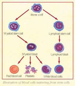
IMAGE: http://www.cancerpictures.us/2009_09_01_archive.html Definition of leukemia: Cancer that starts in blood-forming tissue such as the bone marrow and causes large numbers of blood cells to be produced and enter the bloodstream. Estimated new cases and deaths from leukemia in the United States in 2009: New cases: 44,790 Deaths: 21,870

IMAGE: Swollen, shiny, bleeding gingiva of a patient with acute myelogenous leukemia
http://www.accentu8dental.com.au/page41.php

IMAGE: chronic myeloid leukemia cells
http://www.cancerpictures.us/2009_09_01_archive.html

IMAGE: http://www.cancerpictures.us/2009_09_01_archive.html

IMAGE: A blood smear showing myelogenous leukemia cells (in purple)
http://www.uhnresearch.ca/news/NRX/NRxMay2007.htm

IMAGE: http://www.biologyreference.com/Oc-Ph/Oncogenes-and-Cancer-Cells.html

IMAGE: http://www.stanford.edu/group/cleary/research.html

IMAGE: http://www.topnews.in/drug-inhibits-acute-leukemia-cell-growth-discovered-2156759
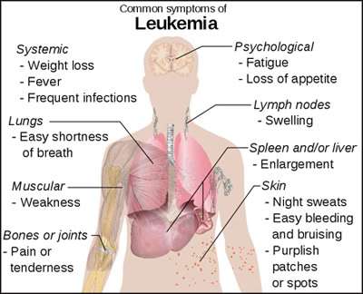
IMAGE: Common symptoms of leukemia

IMAGE: Acute Monocytic Leukemia Symptoms
http://www.livestrong.com/article/113433-acute-monocytic-leukemia-symptoms/
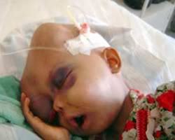
IMAGE: Iraqi child with leukemia
http://www.worldproutassembly.org/archives/2006/05/world_is_blind.html

IMAGE: Marked splenomegaly (swollen spleen):4,000g (chronic myeloid leukemia). A normal spleen of the same age is shown on the right: 110g [Nagasaki City – Peace and Atomic Bomb]
Leukemia
Courtesy of the Research Institute for Radiation Biology and Medicine of Hiroshima University
In the bone marrow in the upper picture, we see various types of young blood cells, including healthy white cells. The marrow below contains only immature leukemic blood cells.

(Courtesy of the Research Institute for Radiation Biology and Medicine of Hiroshima University)
In the bone marrow in the upper picture, we see various types of young blood cells, including healthy white cells. The marrow below contains only immature leukemic blood cells.
Leukemia is cancer of the blood. The white blood cells multiply wildly without fully maturing. Red blood cells and platelets are reduced, leading to anemia. The white blood cells increase in number but lose normal functioning, which lowers resistance to infection. The incidence of leukemia was greatest 7 to 8 years after the bombing. The younger the survivor was at the time of exposure, the earlier was the onset of leukemia.
http://www.pcf.city.hiroshima.jp/virtual/cgi-bin/museum.cgi?no=4028&l=e
Bone marrow displaying acute leukemia

IMAGE: Provided by the Hiroshima Red Cross and Atomic-bomb Survivors Hospital. 1,700m from the hypocenter (Hiratsuka-cho).
A 9-year-old girl exposed in a wooden house received no burns or other injuries. She grew up strong and healthy. In June 1959, about fourteen years after the bombing, she suddenly experienced fatigue, dizziness, bleeding from gums, and other symptoms. Her symptoms persisted, and she was hospitalized. In late June the following year, her legs began swelling without subsiding, eventually to the point of bleeding. She died in late July.
http://www.pcf.city.hiroshima.jp/virtual/cgi-bin/museum.cgi?no=4029&l=e
Age of onset of leukemia
The incidence of leukemia among A-bomb survivors was found to be in proportion to the doses of radiation to which they were exposed. Furthermore, the younger they were when exposed, the higher the leukemia risk. The peak of leukemia onset was about 7 to 8 years after exposure

IMAGE: Source – Effects of A-bomb Radiation on the Human Body 1992,
Ed. Hiroshima International Council for Medical Care of the Radiation-exposed, Bunkodo
http://www.pcf.city.hiroshima.jp/peacesite/English/Stage1/1-5/1-5-6E.html

IMAGE: The younger the person is at the time of exposure, the sooner leukemia develops.
Note: ”High-dose” means1 Sievert or more. Reference: A-bomb Radiation Effects Digest 1992
http://www.pcf.city.hiroshima.jp/virtual/cgi-bin/e/Leukemia_e.htm
Graph of leukemia cases in Hiroshima

http://glasstone.blogspot.com/2007/03/above-3.html
Graph of cancer and leukemia mortality in Hiroshima-Nagasaki

IMAGE: http://glasstone.blogspot.com/2007/03/above-3.html

IMAGE: http://glasstone.blogspot.com/2007/03/above-3.html

IMAGE: Radiation dose to the bone marrow and leukemia. You can see that small doses up to 5 rads have no effect either way on the leukemia risk, while 6-9 rads in this data seems to cause a reduction in normal leukemia risk from 0.17% to 0.12%. Doses which exceed this are harmful, possibly because the P53 repair mechanism was saturated and could not repair radiation induced damage to DNA due to the rate it occurred at higher doses. A dose of 20-40 rads more than doubles the natural leukemia risk. Hence anyone getting leukemia after a larger dose is more than 50% likely to have got the cancer as a result of the radiation exposure than naturally. [The Effects of Nuclear Weapons]
Radiation dose and symptoms

IMAGE: Whole body radiation dose and symptoms. The symptoms that appear after full-body exposure to radiation vary with the dose received. A dose greater than 7,000 millisieverts is nearly 100% lethal. [Hiroshima Peace Memorial Museum]
Body Parts Most Vulnerable to Radiation

IMAGE: The effects of radiation differ with the type of radiation and the parts of the body exposed. Radiation is especially harmful to the blood-forming bone marrow, the mucous membranes in the intestines, the genital organs, and other tissues in which cells divide rapidly.
[Hiroshima Peace Memorial Museum]
Malignant tumors
Beginning around 1960, the incidence of cancer began to increase. The main cancers included thyroid, breast, lung, and salivary gland. The role of radiation in cancer is profoundly significant. Some researchers have reported a direct correspondence between distance from hypocenter, probable exposure dose, and malignancy rates.

IMAGE: Source: Effects of A-bomb Radiation on the Human Body 1992,
Ed. Hiroshima International Council for Medical Care of the Radiation-exposed, Bunkodo
[Hiroshima Peace Memorial Museum]
 IMAGE: Radiation and Radioactivity. Materials that emit “radiation” are called “radioactive materials.” “Radioactivity” refers to the ability to emit radiation. Comparing a radioactive material to a flashlight, for example, the radiation would correspond to the light itself while radioactivity is the ability to emit lights.
[Hiroshima Peace Memorial Museum]
 IMAGE: Donated by Hiroko Yamashita (n e, Yoshida) 800m from the hypocenter Ote-machi About 15 days after the bombing, the mother of Hiroko (then, 18) started to comb her daughter’s hair, but in about three strokes it all came out except for a fringe along her hairline.
At eighteen, Hiroko was exposed in her home 800 meters from the hypocenter. She was seriously injured and trapped under her house. About two weeks after the bombing, she had diarrhea and vomiting and showed other symptoms of radiation poisoning. The purple spots from internal bleeding appeared on her body. One day, all her hair came out with just three swipes of the brush. Hiroko’s mother assumed that Hiroko would not survive, so she saved this hair as a reminder of her daughter. Luckily, Hiroko did recover after fighting the radiation for six months. However, she later developed cancer and suffered both physically and emotionally.
More detail: Hiroko Yamashita (maiden name Yoshida, then, 18) was exposed to the A-bomb in Ote-machi where she was trapped under her collapsed house. She crawled out of the rubble with her younger brother Yusaku (then, 6), and they fled barefoot toward the suburbs from a city bursting into flame. Hiroko’s hands, shoulders, and legs were severely injured. Yusaku, who appeared uninjured, fell ill on the 21st. His hair fell out, he developed a high fever, his nose bled heavily, and he died on the 24th. On the 21st, the day Yusaku’s hair fell out, Hiroko also lost her hair in large clumps. Her condition worsened for some time, but under the devoted care of her mother, Kyo (then,46), she managed to survive. In preparation for the worst, Kyo had kept this hair to treasure as a reminder of Hiroko. [Hiroshima Peace Memorial Museum]

IMAGE: Damage by the Radiation – Acute Effects
Symptoms of radiation poisoning that appeared immediately after the atomic bombing are called acute effects. The unique characteristic of the atomic bombs was the massive amount of radiation they emitted. Conventional bombs emit no such radiation. This radiation severely damaged victims’ bodies. Radiation destroys cells, alters blood quality, damages blood marrow and other blood-forming organs, and causes other serious injury. Anyone directly exposed to radiation within 1,000 meters from thehypocenter received a life-threatening dose, and most died within a few days. Some who appeared completely uninjured began vomiting blood or died with purple spots all over their bodies.
[Hiroshima Peace Memorial Museum]

IMAGE: Damage by the Radiation – aftereffects. The damage done by radiation was not limited to the weeks or months immediately after the bombing. Aftereffects continued to manifest for decades. By the end of 1945, the injuries inflicted by the A-bomb appeared to be healing, but soon keloid scars, cataracts, leukemia, and other cancers began appearing in many survivors. Because Hiroshima was the first atomic bombing in history, no one knew what to expect from exposure to high levels of radiation. Survivors were forced to live with continual anxiety, never knowing what symptoms might appear or when. No effects on descendants of survivors have yet been proven.
[Hiroshima Peace Memorial Museum]

IMAGE: Soldier with Purpura, “Spots of Death” September 3, 1945 This man was exposed in a house within one kilometer of the hypocenter. Ten days after the bombing, his hair began falling out and he bled from the gums. Blood collecting under his skin formed purple spots. His gums bled continuously. He died about one month after the bombing. Photo: Gonichi Kimura

IMAGE: Woman with keloids on the back of her arms and on back.
Keloids and puckered skin caused terrible physical and emotional pain.
October 1945
Courtesy of the US Army
http://www.pcf.city.hiroshima.jp/kids/KPSH_E/hiroshima_e/sadako_e/subcontents_e/13kousyougai_1_e.html
![]()
IMAGE: Close-up of previous photo. [Atomic Bomb Museum]

IMAGE: Provided by the Research Institute for Radiation Biology and Medicine of Hiroshima University 1,000m from the hypocenter A 22-year-old soldier exposed indoors received injuries on his head and back. On August 20, his hair fell out and purpura appeared over his entire body. On September 1, his gums began to swell. On the 3rd he developed cold sores in his mouth and a swollen throat. His high fever persisted. He died at 9:20 a.m. on September 8. His preserved tongue displays black purpura spots in the center. [Hiroshima Peace Memorial Museum]

IMAGE: Firefighter burned at Chernobyl. [RS-500 nuclear radiation detector]
 IMAGE: Chernobyl firefighter with burns and keloids. [Yumi Kikuchi’s blog]

IMAGE: Children of the Atom Bomb (name of site) pilation of Scalp Epilation of the scalp, beginning on the nineteenth day, three days after the appearance of purpura. Scattered long hairs of the original growth remain. Patient was inside a wooden building at Nagasaki. Leukopenia persisted for two months, but the patient recovered. [Children of the Atom Bomb]

IMAGE: Petechiae and purpura. [Dictionnaires et Encyclopédies sur ‘Academic’] Thrombocytopenia or thrombocytosis, caused by a lack of platelets or an abundance of malfunctioning platelets, respectively. Leukemia patients may exhibit increased bruising or bleeding. Signs of this are petechiae, tiny blood spots from capillary hemorraghing, or inexplicable bleeding from seemingly minor injuries. [Leukemia Symptoms]

IMAGE: Breakdown of the A-bomb energy released. [Hiroshima Peace Memorial Museum]

IMAGE: Skin lesions, caused by leukemic cells invading the skin layers. [Leukemia Symptoms]

IMAGE: Hyperplasia of the gums seen in leukemia. [Leukemia Symptoms]
In a healthy human, red blood cells circulate oxygen throughout the body, while white blood cells protect against infection. When a person has leukemia, the malformed leukemic cells replace and block the production of normal cells, causing this system to fail. The most common symptoms of leukemia results from the collapse of the blood’s normal functions. Leukemia patients often experience a variety of indeterminate symptoms, such as weight loss or joint pain, before their diagnosis. The symptoms of leukemia include:
*Anemia, caused by a lack of red blood cells. Patients tend to suffer from weakness, fatigue, and malaise, and their skin may look pale from anemia. When exercising, anemic patients may feel faint or experience shortness of breath.
*Leukopenia, caused by a lack of normal white blood cells. Patients show increased susceptibility to infection. They may be slow to heal from minor injuries or experience skin or organ infections.
*Neutropenia, caused by a lack of mature neutrophils.
*Thrombocytopenia or thrombocytosis, caused by a lack of platelets or an abundance of malfunctioning platelets, respectively. Leukemia patients may exhibit increased bruising or bleeding. Signs of this are petechiae, tiny blood spots from capillary hemorraghing, or inexplicable bleeding from seemingly minor injuries.
*Enlarged lymph nodes, spleen, or liver, caused by the accumulation of leukemic cells.
*Skin lesions, caused by leukemic cells invading the skin layers.
Mesothelioma
Mesothelioma is usually associated with asbestos exposure but 20% of cases are thought to be caused by radiation. [REF]
There is a notably high incidence of mesothelioma (highly statistically significant based on 3 patients). This cancer is usually associated with exposure to asbestos but 20% of cases are thought to be due to radiation, according to the Oxford Textbook of Pathology.
Pathopshysiology of lung damage by inhaled radionuclides
The pathophsyiology of lung damage caused by inhaled respiratory radionuclides is described thus [REF]:
The inhaled particle tracks down through the respiratory passages into the lungs, settling in an alveolar sac. The radioactive particle emits radioactive rays, alpha, beta and gamma rays that affect the cells nearby, damaging the DNA and causing malignant transformation. The damaged cell reproduces producing a colony of rapidly dividing cells that eventually out-multiply normal cells, and ultimately cause death of the organism.
In this picture, a plutonium particle has lodged in the lung of an ape. Tracking the particle for 48 hours, this composite picture tracks the path of the alpha rays emanating from the particle. These rays cause chromosomal abnormalities in neighboring cells and cause cancer to develop.
Alpha Rays from a Radioactive Particle in Lung Tissue

IMAGE: The black star shows the tracks made over a 48 hour period by alpha rays emitted from a radioactive particle of plutonium lodged in the lung tissue of an ape (the particle itself is invisible). In living lung tissue, if one of the cells adjacent to the particle is damaged in a certain way, it can become a cancer cell later on, spreading rapidly through the lung, causing almost certain death.
Photo by Robert Del Tredici from his book entitled At Work In The Fields Of The Bomb (Harper and Row, 1987)
Radiation dose and cancer
Below is a table which has been adapted from Answers.com Radiation Poisoning.
Table of exposure levels and symptoms
Dose-equivalents are presently stated in sieverts:
| 0.05–0.2 Sv (5–20 REM) | No symptoms. Potential for cancer and mutation of genetic material although some say this dose is protective against cancer (hormesis) |
| 0.2–0.5 Sv (20–50 REM) | No noticeable symptoms. Red blood cell count decreases temporarily. |
| 0.5–1 Sv (50–100 REM) | Mild radiation sickness with headache and increased risk of infection due to disruption of immunity cells. Temporary male sterility is possible. |
| 1–2 Sv (100–200 REM) | Light radiation poisoning, 10% fatality after 30 days. Typical symptoms include mild to moderate nausea, with occasional vomiting, beginning 3 to 6 hours after irradiation and lasting for up to one day. This is followed by a 10 to 14 day latent phase, after which light symptoms like general illness and fatigue appear. The immune system is depressed, with convalescence extended and increased risk of infection. Temporary male sterility is common. Spontaneous abortion or stillbirth will occur in pregnant women. |
| 2–3 Sv (200–300 REM) | Severe radiation poisoning, 35% fatality after 30 days. Nausea is common, with 50% risk of vomiting at 2.8 Sv. Symptoms onset at 1 to 6 hours after irradiation and last for 1 to 2 days. After that, there is a 7 to 14 day latent phase, after which the following symptoms appear: loss of hair all over the body, fatigue and general illness. There is a massive loss of leukocytes (white blood cells), greatly increasing the risk of infection. Permanent female sterility is possible. Convalescence takes one to several months. |
| 3–4 Sv (300–400 REM) | Severe radiation poisoning, 50% fatality after 30 days. Other symptoms are similar to the 2–3 Sv dose, with uncontrollable bleeding in the mouth, under the skin and in the kidneys after the latent phase. |
| 4–6 Sv (400–600 REM) | Acute radiation poisoning, 60% fatality after 30 days. Fatality increases from 60% at 4.5 Sv to 90% at 6 Sv (unless there is intense medical care). Symptoms start half an hour to two hours after irradiation and last for up to 2 days. After that, there is a 7 to 14 day latent phase, after which generally the same symptoms appear as with 3-4 Sv irradiation, with increased intensity. Female sterility is common at this point. Convalescence takes several months to a year. The primary causes of death (in general 2 to 12 weeks after irradiation) are infections and internal bleeding. |
| 6–10 Sv (600–1,000 REM) | Acute radiation poisoning, near 100% fatality after 14 days. Survival depends on intense medical care. Bone marrow is nearly or completely destroyed, so a bone marrow transplant is required. Gastric and intestinal tissue are severely damaged. Symptoms start 15 to 30 minutes after irradiation and last for up to 2 days. Subsequently, there is a 5 to 10 day latent phase, after which the person dies of infection or internal bleeding. Recovery would take several years and probably would never be complete.Devair Alves Ferreira received a dose of approximately 7.0 Sv (700 REM) during the Goiânia accident and survived, partially due to his fractionated exposure. |
| 10–50 Sv (1,000–5,000 REM) |
The mouth of a man who has suffered a 10 to 20 Gy dose 21 days after the exposure, note that damage to normal skin, the lips and the tongue can be seen Acute radiation poisoning, 100% fatality after 7 days. An exposure this high leads to spontaneous symptoms after 5 to 30 minutes. After powerful fatigue and immediate nausea caused by direct activation of chemical receptors in the brain by the irradiation, there is a period of several days of comparative well-being, called the latent (or “walking ghost“) phase. After that, cell death in the gastric and intestinal tissue, causing massive diarrhoea, intestinal bleeding and loss of water, leads to water-electrolyte imbalance. Death sets in with delirium and coma due to breakdown of circulation. Death is currently inevitable; the only treatment that can be offered is pain therapy. Louis Slotin was exposed to approximately 21 Sv in a criticality accident on 21 May 1946, and died nine days later on 30 May. At this dose the skin can be damaged, here is a photo of a man who received a 10 to 20 Gy gamma whole body dose as a result of an industrial accident, He died about 10 days after the photo was taken, about 30 days after the event. |
| 50–80 Sv (5,000–8,000 REM) | Immediate disorientation and coma in seconds or minutes. Death occurs after a few hours by total collapse of nervous system. |
| More than 80 Sv (>8,000 REM) | U.S. military forces expect immediate death. A worker receiving 100 Sv (10,000 REM) in an accident at Wood River, Rhode Island, USA on 24 July 1964 survived for 49 hours after exposure, and an operator receiving between 60 and 180 Sv (18,000 REM) to his upper body in an accident at Los Alamos, New Mexico, USA on 30 December 1958 survived for 36 hours; details of this accident can be found on page 16 (page 30 in the PDF version) of Los Alamos’ 2000 Review of Criticality Accidents. |

Types of radiation
 IMAGE: Radiation types and their penetrating power. [Hiroshima Peace Memorial Museum]
Breakdown of Energy Released

http://www.pcf.city.hiroshima.jp/peacesite/English/Stage1/1-4/1-4-4E.html
Fallout
Black rain
“Black rain”
The Hiroshima and Nagasaki explosions yielded some 200 different kinds of radioactive isotopes, that is, nuclear fission particles of uranium and plutonium that escaped fission. Following the explosions, these and other materials irradiated by neutrons from the bomb, were carried high into the atmosphere.
The mixing of enormous amounts of airborne irradiated materials combined with heat and thermal currents from the firestorms led to rainfall in both cities within 30-40 minutes of the bombings. As the fallout particles were mixed with carbon residue from citywide fires, the result was the awesome—and injurious—“black rain.”
This “black rain” reached ground level as sticky, dark, dangerously radioactive water. It not only stained skin, clothing, and buildings, but also was ingested by breathing and by consumption of contaminated food or water, causing radiation poisoning.
 IMAGE: Atmospheric dispersion begins in Hiroshima [Atomic Bomb Museum]
 IMAGE: Donated by Akijiro Yajima 3,700m from the hypocenter Furuta-machi, Takasu The roof of a house 3,700 meters from the hypocenter was dislodged by the blast, allowing in drops of black rain that dripped down and left traces on this white plaster wall. Analysis of this rain revealed that it contained radioactive fallout from the atomic bomb explosion. [Hiroshima Peace Memorial Museum]
 IMAGE: Sketches made immediately after the atomic bomb exploded
These sketches, illustrating the first 30 minutes after the explosion, were drawn by meteorologists at what was then the Hiroshima District Meteorological Observatory.
Dirt, soot, and other matter flung up from the ground surface formed black smoke. That dirt and soot mixed with water drops in the air and formed a black rain that fell. [Hiroshima Peace Memorial Museum]

IMAGE: Shirt stained by black rain. Donated by Tokuso Wakamoto. 1800m from the hypocenter. Fukushima-cho. Tokuso Wakamoto (then 28) was exposed to the bomb on the road en route to his assigned building demolition site. While fleeing the city, he was caught in black rain in western area of city (about 3,500 meters from hypocenter). The sticky black rain left black stains on his clothing. [Hiroshima Peace Memorial Museum]
Residual radiation
Radioactivity: two kinds – initial radiation (explained above), and induced radiation (also called “residual radiation”).
The radioactive material used in the Hiroshima bomb was uranium. Of the approximately 50 kilograms of uranium packed into the bomb, only about one kilogram underwent fission. About 15% of the energy released was in the form of radiation. The radiation released the instant the nuclear fission took place is called “initial radiation.” The large amounts of radiation remaining on the surface for some time after the explosion is called “residual radiation.”

IMAGE: Effects of Residual Radiation Residual radiation had devastating effects on human bodies. However, this residual radiation faded rapidly. A week later, it was about one millionth of the original level. Today, residual radiation from the Hiroshima A-bomb has no effect on human bodies. [Hiroshima Peace Memorial Museum]
2. Initial radiation from the bombs
About 3% of the Hiroshima and Nagasaki bombs’ energy was spent in generating ionizing radiation—high-energy particles and rays with enough energy to “ionize” neutral atoms, i.e., strip electrons away from them. Some of this ionized radiation was absorbed by the air, but neutrons (electrically neutral sub-atomic particles) and gamma and X-rays (extremely high energy forms of light) did reach the ground, and these rays damaged exposed living tissues. Close to ground zero of both explosions, dosages were high enough to be immediately lethal for persons not already killed by flash, blast, or fire.
3. Induced radioactivity
Initial bursts of radiation from the two bombs also created induced or residual radioactivity. Soil and other materials were irradiated in the blast areas. Absorption of “slow neutrons” by all kinds of substances caused the creation of new isotopes that then emitted ionizing radiation.
Japanese physicists examining the areas near ground zero in Hiroshima found unusually high levels of radioactivity in the soil, in the bones of a horse, and even in the sulphur content of electrical insulators on telephone poles. Eventually, a variety of unusual radioactive elements were found in soil, roofing tiles, asphalt, and concrete near ground zero in the two cities. There were many instances of radiation effects on animals and plants.
|
Age
|
Sex
|
Symptoms
|
Diagnosed Illness
|
Time Spent at Ground Zero
|
| 46 |
M
|
– Burning of esophagus | – Acid reflux – Hiatal hernia (life long) |
Lab (10 yrs.) Morgue (2 weeks) WTC (1 week) Drove CO back and forth with evidence |
| 35 |
M
|
– Persistent cough – Chest pain – Decreased immune system |
– Sarcoidosis – Asthma – Rheumatoid arthritis |
WTC (120 hours) started working on the 3rd day |
| 52 |
M
|
– Throat cancer | ||
| 49 |
M
|
– No symptoms | – Aortic aneurysm | Ground Zero (1 Month) |
| 37 |
F
|
– Shortness of breath | – Sarcoidosis | – Clean-up (bucket brigade) – Security at WTC (4 months) |
| 42 |
M
|
– Shortness of breath | – Reactive airways dysfunction syndrome (occupational asthma) | WTC (between 400 and 500 hours) |
| 33 |
M
|
– Chronic cough | – Post-traumatic stress | – WTC (1 day) – Fresh Kills (1 day) |
| 40 |
F
|
– No symptoms | – Rectal cancer Sep. 2002 | Security at ground zero (1 month) |
| 48 |
M
|
– No symptoms – Blood in urine |
– Renal cell cancer (kidney cancer) | WTC – security ( 2 weeks) |
| 37 |
M
|
– Extended cough | – Sarcoidosis | WTC (2 weeks) – security and traffic control |
| 43 |
M
|
– Bile-duct cancer Deceased |
First Responder – WTC (6 months) | |
| 48 |
M
|
– Tightness in chest – High blood pressure |
Security in hot zone (30 days) | |
| 33 |
M
|
– Headaches | – Brain cancer | WTC (2 weeks) Bucket brigade, security |
| 35 |
M
|
– Lump on neck | – Thyroid cancer – Stage 2 multiple myeloma |
WTC (12 days) S.I. landfill (61 days) |
| 34 |
M
|
– Difficulty breathing | – Pulmonary disease – Respiratory failure Deceased |
WTC (3 months) |
| 47 |
M
|
– Shortness of breath – Joint stiffness |
– Heart attack – Sarcoidosis Deceased |
WTC (3 months) |
| 39 |
F
|
– No symptoms | – Sarcoidosis | WTC (1 month) |
| 42 |
M
|
– Bronchitis (3 times) – Pneumonia – Shortness of breath – Loss of intake |
– Chronic lung disease | Morgue (9 months) |
| 38 |
F
|
– Joint pain – Shortness of breath – Chest pain |
– Sarcoidosis | WTC (5 days) |
| 48 |
M
|
– Swelling of hands – Shortness of breath – High hemoglobin level |
– Scarred lung tissue | WTC (2 days) S.I. landfill (1 day) |
| 41 |
M
|
– Irregular heartbeat – Shortness of breath – Eyes were burned |
– Tumor in left lung – Debris, scarring, loss of function in right lung – Cataracts |
South tower on 9-11 WTC (1 year—periodically) |
| 34 |
M
|
– Difficulty breathing – Tightness in chest. |
– Blocked airway | WTC (5 days at 12 hours) |
| 41 |
M
|
– Mass in abdomen – Trouble breathing – Skin rashes |
– Lymphoma | WTC (3 months) |
| 32 |
M
|
– Shortness of breath – chest pains |
– Asthma | S.I. Landfill (1 week) WTC (1 week) |
| 44 |
M
|
– Weight loss – shortness of breath – loss of appetite – dry mouth |
– Pulmonary sarcoidosis | WTC (30 days) |
| 44 |
M
|
– Coughing blood and – shortness of breath |
– BOOP [bronchiolitis obliterans organizing pneumonia] – Bronchitis |
WTC (Start to finish) First responder |
| 36 |
M
|
– Coughing – shortness of breath – wheezing – extreme heartburn |
– Asthma – RADS [reactive airways disease] – Sleep apnea |
WTC (4 months) |
| 39 |
M
|
– Difficulty breathing – Fatigue – Increased sensibility to heat |
– Pulmonary restricted lung disease | WTC (1 week) S.I. Landfill (1 week) First Responder |
| 45 |
M
|
– Anxiety | – Hypertension | WTC (15 days) |
| 42 |
M
|
– Difficulty breathing – Fatigue |
– Prostate cancer – Sleep apnea – Restrictive lung disease |
WTC (4 months) First Responder |
| 42 |
M
|
– Shortness of breath – Coughing |
– Chronic obstructive pulmonary disease – Asthma |
WTC (6 months) |
| 34 |
M
|
– Difficulty breathing – Fatigue – Anxiety |
– Decreased airway – High blood pressure |
WTC (3 months) |
| 40 |
M
|
– Coughing up blood – Persistent cough and wheezing – Gasping |
– Cardiomyopathy – Chronic heart failure |
WTC (3 months) First Responder |
| 45 |
F
|
– Swelling of the joints – Pain in the bones, hips – Tumors |
– Sarcoidosis | WTC (6 weeks) |
| 34 |
M
|
– Vomiting – Difficulty breathing – Chest pains – Diarrhea – Dizzy spells |
– Asthma – GERD [Gastro-esophageal reflux disease] – Post-traumatic stress – Chemical induced bronchitis – Acid reflux |
WTC (3 weeks ) First Responder |
| 37 |
M
|
– Colds – Difficulty breathing – Winded – Fatigue |
– Chronic sinusitis | WTC S.I. Landfill (1 month and half) |
| 30 |
F
|
– Swelling of the joints – Swelling of the lymphoid – Numbness of arms and legs |
– Sarcoidosis | WTC – S.I. Landfill and Morgue (5 weeks) |
| 40 |
M
|
– Difficulty breathing | – Chronic sinusitis | WTC (6 months) |
| 42 |
M
|
– Difficulty breathing | – Chronic sinusitis – Diminished lung capacity – Reflux |
WTC (9 Months) |
| 47 |
M
|
– Relatively no symptoms – Coughing |
– Heart attack – Triple bypass – Advanced throat cancer – Acid reflux |
WTC (3 months) First Responder |
| 46 |
M
|
– Upper respiratory infection – Shortness of breath |
– Acid reflux – GERD – Esophagitis – Reactive airways disease – Dyspnea – Extra thoracic airways disease – Sleep apnea – Seizures |
WTC (4 months) |
| 40 |
M
|
– Difficulty breathing – Fatigue – Wheezing |
– Bronchitis | WTC (4 months) |
| 38 |
F
|
– Abdominal pain – Heavy bleeding – Fatigue |
– Adenoid carcinoma – Carcinoid (stomach, gall bladder, uterus, fallopian tube, ovaries, appendix and colon) |
WTC (2 months) First Responder |
| 39 |
M
|
– Shortness of breath | – Decreased lung capacity | WTC (3 months) First Responder |
| 44 |
M
|
– Jaundice – Fatigue |
– Body is destroying red blood cells. Dr. wants to remove his spleen. ( No diagnosis.) | WTC Morgue (6 months) First Responder |
| 48 |
M
|
– Anxiety – Headache – Difficulty breathing – Dizziness |
– Polypoid sinus disease – Asthma – Lung disease – Acid reflux |
WTC (6 months) 1 year sifting through remains First Responder |
| 36 |
F
|
– Chronic fatigue – Trouble breathing – Swollen neck |
– Thyroid cancer | WTC (5 months) First Responder |
| 48 |
M
|
– Severe cough | – Nasal polyps – Asthma |
WTC (3 months) |
| 46 |
M
|
– Cough – Shortness of breath – Phlegm – Wheezing and – Difficulty sleeping |
– No diagnosis | Morgue (3 months) First responder |
| 55 |
M
|
– Trouble sleeping – Runny nose |
– Post-traumatic stress syndrome – Sinusitis |
WTC (1 year) First Responder |
| 45 |
M
|
– Shortness of breath – Chest pain – Excessive protein in urine |
– Sarcoidosis – Nephritic syndrome (affects the kidney) |
WTC (3 months) First Responder |
| 40 |
M
|
– Jaundice – Fatigue – Shortness of breath – Throwing up blood – Joint pains – Swelling of limbs – Blurred vision – Numbness in finger tips and toes |
– Failed liver non-alcoholic steatohepatitis (liver transplant) – GERD |
WTC (1 month and a half) First Responder |
| 49 |
M
|
– Headaches – Coughing – Fatigue |
– Sinusitis | WTC (1 month) |
| 69 |
M
|
– Runny nose – Lung congestion |
– Sinusitis – Pre-cancerous kidney (removed) |
WTC (3weeks) |
| 43 |
M
|
– Shortness of breath – Chronic rashes – Joint pain – Numbing of limbs – Tumors |
– Sarcoidosis | WTC (2 months) S.I. Landfill (2 months) Morgue (15 days) |
| 45 |
M
|
– Pain in left side – High calcium level – Fatigue – Dizziness |
– Kidney stones | WTC (1 month) First Responder |
| 29 |
M
|
– Coughing up blood and mucus | – Chronic upper respiratory infection | WTC (2 weeks) |
| 42 |
F
|
– Coughing – Eye irritation – Headaches |
– Sinusitis | WTC (1 month and a half) First Responder |
| 43 |
M
|
– Loss of hearing – Runny nose – Difficulty breathing – Heartburn |
– Loss of hearing | WTC (9 months) First Responder |
| 36 |
M
|
– Nose bleed – Sinus infection – Difficulty sleeping – WTC cough |
– Post traumatic stress disorder – Hypertension – Kidney stones – Sinusitis |
WTC (2 weeks) First Responder |
| 47 |
M
|
– Shortness of breath – Irregular heartbeat – Wheezing – Fatigue – Numbness |
– Sleep apnea | WTC (3 days) First Responder Volunteer |
| 47 |
M
|
– Difficulty sleeping – Tightness in chest – Neckaches – Bad cough – Headache |
– Acid Reflux – Reactive airways |
WTC (2 months) First Responder |
| 38 |
M
|
– Difficulty breathing – Constant infections – Fatigue |
– Thyroid cancer – Chronic sinusitis |
WTC (3 months) First Responder |
| 32 |
M
|
– Fatigue – Shortness of breath – Migraine headaches – Coughing |
– RAD | WTC (3 months) First Responder |
| 36 |
M
|
– Chronic cough – Fatigue |
– Acid reflux | WTC (1 week) First Responder |
| 44 |
M
|
– Difficulty breathing – Short- tempered – Runny nose |
– Sinusitis – Acid reflux |
WTC (18 months all together) Morgue S.I. landfill First Responder |
| 44 |
M
|
– Heartburn – Loss of appetite |
– Acid reflux – Hiatal hernia – Irritation to stomach lining |
WTC (6 months) First Responder |
| 35 |
M
|
– Shortness of breath | WTC (2 months) | |
| 35 |
M
|
– Protein in urine – High cholesterol – Dizziness – Fatigue – Swelling |
– Focal segmental glomerular sclerosis (kidneys) | WTC (6 months) First Responder |
| 37 |
M
|
– Headaches – Fatigue – Runny nose |
– Occupational asthma – High liver count – Acid reflux – Sinusitis |
WTC (3 months) |
| 40 |
M
|
– Shortness of breath – Fatigue – Difficulty sleeping |
– RADS | WTC (6 months) First Responder |
| 58 |
M
|
– Mass on right side of neck – Unusual snoring – Blood in the mucus |
– Tonsil cancer | WTC (1 week) First Responder |
| 35 |
F
|
– Allergies – Excessive bowl movement – Abdominal pain – Dry cough |
– Crohn’s disease – Fistula |
|
| 46 |
M
|
– Upper respiratory infection | – Crohn’s disease – Allergies |
WTC |
| 48 |
M
|
– Diabetes – Hip replacement – High blood pressure |
– Diabetes – Hip replacement – High blood pressure |
WTC (1 ½ months) First Responder |
| 38 |
M
|
– Severe headache | – Brain tumor – Liver tumor |
WTC (4 months) First Responder |
| 34 |
M
|
– Low lung volume | WTC (2 weeks) First Responder |
|
| 49 |
M
|
– Cough – Difficulty sleeping – Congestion |
WTC (2 weeks) | |
| 42 |
M
|
– Shortness of breath | – Asthma – Acid reflux – Bronchitis |
WTC (3 months) First Responder |
| 43 |
M
|
– Shortness of breath – Fatigue – Moodiness – Nosebleeds |
– Nodules on lungs – Acid reflux |
WTC (6 months) First Responder |
| 35 |
M
|
– Diarrhea – Blood in urine |
– Colitis | WTC (5 months) First Responder |
| 43 |
M
|
– Blood in urine – Acid reflux |
– Bladder cancer | WTC (1 week) |
| 46 |
M
|
– Anxiety – Difficulty sleeping – Runny nose – Tightness in the chest |
– Sinusitis – Sleep apnea – Post traumatic stress |
WTC (4 months) First Responder |
| 43 |
M
|
– Wheezing – Shortness of breath – Joint pain |
WTC (3 weeks) First Responder |
|
| 27 |
M
|
– Chronic cough – Difficulty breathing – Upper respiratory infection |
– RADS – Sinusitis |
WTC (3 weeks) First Responder |
| 38 |
M
|
– Shortness of breath – Coughing – Fatigue – Aches – Headaches – Loss of appetite |
– Sarcoidosis | WTC (3 months) First Responder |
| 36 |
M
|
– Shortness of breath – Dizziness – Headache – Vertigo – Joint pain |
– Sarcoidosis | WTC (approx. 4 months) First Responder |
| 49 |
F
|
– Scar tissue on the lungs – Shortness of breath – Fatigue – Joint pain |
– Obstructive pulmonary disease | WTC (2 months) First Responder |
| 46 |
M
|
– Rash – Headaches – Shortness of breath – Joint pain – Short temper – Anxiety – Difficulty sleeping |
– Acid reflux – High mercury count |
WTC (1 month) First Responder |
| 45 |
M
|
– Stomach aches – Low red blood cell count – Low white blood cell count |
– Spleen removed | WTC (1 week) First Responder |
| 42 |
F
|
– Recurring cyst | WTC (3 months) First Responder |
|
| 39 |
M
|
– Difficulty sleeping – Anxiety |
– Post traumatic stress disorder | WTC (7 months) First Responder |
| 45 |
M
|
– Fatigue – Difficulty breathing – Joint pains vertigo – Headaches – Anxiety |
– Sinusitis – Acid reflux – Ulcer |
WTC (6 months) First Responder |
| 44 |
F
|
– Difficulty breathing – Difficulty sleeping |
– RADS – Vocal cord damage |
WTC (3 months) First Responder |
| 43 |
F
|
– Bronchitis – Acid reflux – Sinusitis – Cough |
– Sarcoidosis | WTC (3 months) First Responder |
| 44 |
M
|
– Cough – Difficulty breathing |
– RADS – Sinusitis – Rhinitis – Esophagitis – GERD – Sleep apnea – Lung scarring |
WTC (2 months) S.I. Landfill (1 month |
| 37 |
M
|
– Anxiety – Depression |
– Post traumatic stress disorder | WTC First Responder |
| 31 |
M
|
– Diarrhea – Blood in stool |
– Colitis | WTC (2 weeks) First Responder |
| 49 |
M
|
– Blood in urine – Difficulty breathing |
– Tumor in the bladder | WTC (4 months) First Responder |
| 44 |
M
|
– Difficulty breathing – Joint pain – Recurring bronchitis |
– Sarcoidosis – Asthma |
WTC (1 month) First Responder |
| 38 |
M
|
– Fatigue – Headaches – High blood pressure – Difficulty breathing |
– Glomerulo-nephritis (kidney disease) | WTC (2 weeks) First Responder |
| 35 |
M
|
– Bloating – Heartburn – Loss of appetite |
– Acid reflux | WTC (6 months) Morgue (2 months) |
| 41 |
M
|
– Bone pain – Back pain |
– Multiple myeloma (cancer of the plasma cell) | WTC (2 days) Morgue & S. I. Landfill (3 weeks) |
| 44 |
M
|
– Lump in the leg | – Soft tissue cancer | WTC (1 week) |
| 45 |
F
|
– Joint pain – Trouble sleeping |
– Leukemia | WTC (2 years) |
| 47 |
M
|
– Lump in throat | – Throat cancer | WTC (2 ½ months) |
| 47 |
M
|
– Muscle pain | – Herniated disk – Partial tears in muscle tissue |
WTC (2 weeks) S.I. Landfill (1 week) |
| 37 |
F
|
– Shortness of breath – Tightness of the chest |
– Asthma – Sleep apnea |
WTC (2 months) |
| 44 |
M
|
– Difficulty breathing | – Heart attack | WTC (2 months) First Responder |
| 46 |
M
|
– Shortness of breath | – Lung cancer | WTC & Morgue (8 months) |
| 49 |
M
|
– Shortness of breath – Difficulty sleeping – Coughing |
– PTSD – Restricted lung disease |
WTC (8 months) |
| 43 |
M
|
– Severe cough | – Asthma | WTC (1 ½ months) |
| 46 |
M
|
– Stomach pain – Difficulty breathing |
– Acid reflux – High mercury count |
WTC (1 month) First Responder |
| 46 |
M
|
– Headaches – Severe cough – Difficulty sleeping – Short tempered – Fatigue – Joint pain |
– High blood pressure – Diabetes – Spots on the lungs – PTSD – Removal of gall bladder (impacted due to stones) |
WTC (9 months) First Responder |
| 38 |
M
|
– Swollen ankles – Chronic cough – Night sweats |
– Sarcoidosis | WTC (2 weeks) First Responder |
| 32 |
M
|
– Lumps – Anal fistulas – Hernia – Acid reflux – Shortness of breath – Skin rashes |
– Crohn’s disease | WTC (2 1/2 months) First Responder |
| 39 |
M
|
– Difficulty breathing and sleeping | – Asthma – PTSD |
WTC (3 months) |
| 40 |
M
|
– Shakes – Difficulty sleeping – Severe cough |
– High blood pressure (still in testing phase) | WTC (2 1/2 months) First Responder |
| 37 |
M
|
– Rash | – Melanoma | WTC (3 months) First Responder |
| 47 |
M
|
– Frequent migraines – Difficulty sleeping |
– Glaucoma | WTC (2 months) First Responder |
| 40 |
M
|
– Shortness of breath | – Asthma | WTC (2 months) First Responder |
| 44 |
M
|
– Bloating – Weakness – Difficulty eating and drinking – Dehydration |
– Short bowel syndrome (large part of small intestine removed) | WTC (2 months) First Responder |
| 46 |
M
|
– Headaches – Backaches |
– Polycythemia vera (clonal stem cell disorder) | WTC (3 months) First Responder |
| 52 |
M
|
– Difficulty breathing – Wheezing |
– Asthma | WTC First Responder |
| 48 |
M
|
– Difficulty breathing | – Asthma | WTC (1 1/2 months) First Responder |
| 30 |
M
|
– High liver enzymes – Gastritis – Heartburn – Nausea – Stomach pain |
– Liver biopsy/colonoscopy – Gall bladder removed |
WTC (4 months) First Responder |
| 40 |
M
|
– Back pain – Night sweats – Difficulty sleeping |
– Cancer of the kidneys and liver | WTC First Responder |
| 33 |
M
|
– Bronchitis – Chest pain – Migraines – Cough – Difficulty breathing |
– Duodenal polyps – GERD – Reflux – Two nodules in the lungs – Sleep apnea – Sinusitis |
WTC (9 months) First Responder |
| 38 |
M
|
– Difficulty breathing – Fatigue |
– Rotting trachea – Decreased lung capacity |
WTC (9 months) First Responder |
| 49 |
M
|
– Congestion – Headaches – Shortness of breath – Loss of sight |
– Adenocarcinoma (cancer in the left sinus) Deceased (2004) |
WTC |
| 36 |
M
|
– Cough – Fatigue – Weight loss – Reduced lung capacity – Irregular heart beat |
– Sarcoidosis | WTC (2 months) |
| 48 |
M
|
– Shortness of breath – Chronic bronchitis – Stiffness in the chest – Difficulty sleeping |
– Asthma – RADS – Sleep apnea |
WTC First Responder |
| 52 |
M
|
– Shortness of breath – Difficulty breathing |
– Pulmonary fibrosis | WTC (4 months) |
| 52 |
M
|
– Lump in the throat – Difficulty sleeping – Difficulty breathing – Vomiting |
– pre-cancer of the esophagus – gastroparesis (hardening of stomach muscle) – GERD |
WTC First Responder |
| 43 |
M
|
– Shortness of breath – Wheezing – Cough |
– Asthma – GERD |
WTC (6 months) |
| 38 |
F
|
– Shortness of breath – High blood pressure – Chest pain – Difficulty sleeping – Joint pain |
– RADS – Sleep Apnea – GERD – Vocal cord dysfunction – Sinusitis – Enlarged heart – Decreased lung capacity – Nodules in the lung |
WTC |
| 43 |
M
|
– Wheezing – Tightness in the chest |
– Asthma | WTC (6 months) |
| 52 |
M
|
– Shortness of breath – Bronchitis – Muscle pain |
– Acid reflux – Heart attack (2002) |
WTC First Responder |
| 36 |
F
|
– Difficulty breathing – Fatigue – Headaches |
– Asthma – Thyroid issues (not yet diagnosed) |
WTC First Responder |
| 43 |
M
|
– Constant headache – Dizziness – Earaches – Difficulty breathing – Joint pains – Difficulty sleeping |
– Sinusitis – Chronic bronchitis |
WTC First Responder |
| 49 |
M
|
– Earaches – Nodules on the lung – Difficulty sleeping – Muscle aches |
– Inflamed lymph nodes – Chronic conjunctivitis – Calcified heart – Calcified aortic valve |
WTC First Responder (1½ months) |
| 48 |
M
|
– Throat burning – Chronic cough – Chest pain – Difficulty sleeping – Headaches – Dizziness – Memory loss |
– PTSD – Asthma – Acid Reflux |
WTC First Responder |
| 45 |
F
|
– Difficulty swallowing | – Neck tumor | WTC First Responder (7 months) |
| 36 |
M
|
– Vertigo – Inability to maintain balance |
– Tumor in pituitary gland | WTC (2 weeks) |
|
M
|
– Post nasal drip – Difficulty breathing |
– Sarcoidosis | WTC (3 months) | |
| 37 |
M
|
– Stuffy nose – Headaches – Sore throat – Wheezing |
– Acid reflux – Sinusitis |
WTC (6 months) |
| 44 |
M
|
– Chronic cough – Difficulty breathing |
– RADS | WTC First Responder |
| 42 |
M
|
– Night sweats – Difficulty breathing |
– RADS – Lymphatic tumors in the chest– Asbestos exposure in the lung (precursor to cancer of the lung)– GERD |
WTC First Responder |
| 33 |
M
|
– Acid reflux – Lung infection – Swelling in the throat – Nasal and sinus problem – Sleep apnea |
– Tonsil, uvula, soft palette, septum removed (genioglossal advancement) – Pneumonia – Chronic sinusitis – Asthma – Sinusitis |
WTC (4 months) First Responder |
| 46 |
M
|
– Difficulty breathing – Headaches – Passing out – Imbalance – Memory loss – Vertigo |
– Diffuse large B cell lymphoma (cancer of the lymph nodes) | WTC First Responder |
| 48 |
M
|
– Difficulty breathing – Headaches – Passing out – Imbalance – Memory loss – Vertigo |
– RADS – Acid Reflux – Vestibular dysfunction (brain damage) – Gait ataxia (tumor) below ribs |
WTC & S.I. (8 months) First Responder |
| 35 |
M
|
– Shortness of breath – Dizziness spells – Swelling of the lungs – Joint pains – Headaches – Constant lung infections |
– Asthma | WTC & S.I. (1 year) First Responder |
| 38 |
M
|
– Persistent cough – Difficulty breathing – Headaches – Lung infection – Scarring on the lungs |
– Asthma – Sinusitis – RADS |
WTC & S.I. (1 year) First Responder |
| 31 |
M
|
– Persistent stuffy nose – Chest pains |
– RADS | WTC (2 years) First Responder |
| 45 |
M
|
– Dizziness – Vertigo – Blurry vision – Imbalance – Headaches |
– Benign positional vertigo | WTC & S.I. First Responder |
| 36 |
F
|
– Wheezing – Aching bones – Repeated bronchitis – Headache |
– Asthma | WTC (2 months) |
| 35 |
M
|
– Pains in the chest | – Esophagitis | WTC (1 week) |
| 41 |
M
|
– Headaches – Sinus infections – Stomach pains – Wheezing – Difficulty sleeping |
– Sinusitis – Erosion of the esophagus – Acid Reflux – Recurring stye in the eye |
WTC First Responder |
| 54 |
M
|
– Bleeding polyps – Heartburn – Stomach pains |
– Colon cancer | WTC (1 month) |
| 35 |
M
|
– Numbness in hands and feet – Peeling hands – Sore throat – Tonsillitis – Deviated septum |
– Allergic rhinitis | WTC First Responder |
| 48 |
F
|
– Stomach pains | – Colon rectal cancer | WTC (2 weeks) |
| 38 |
M
|
– Difficulty breathing – Blurred vision |
– Sinusitis – Scarring of the retina |
WTC First Responder |
| 45 |
F
|
– Lump in left breast | – Breast cancer | WTC First Responder |
| 43 |
F
|
– Headaches – Joint pains – Difficulty sleeping |
– No diagnosis | WTC First Responder |
| 48 |
M
|
– No symptoms | – Renal cell carcinoma (kidney cancer) | WTC |
| 47 |
M
|
– Cough | – Heart attack (deceased) | WTC First Responder |
| 50 |
M
|
– Fatigue – Runny nose |
– Barrett’s esophagus (pre-cancer of the esophagus) | WTC First Responder |
| 35 |
M
|
– Fatigue – Shortness of breath – Nose bleeds – Joint pains |
– Chronic sinusitis – Ground glass in the lungs – Pulmonary hypertension |
WTC First Responder |
| 28 |
M
|
– Cough | – Lymphoma | WTC First Responder |
| 38 | F | – Difficulty breathing – Blurry vision – Joint pain – Memory loss – Difficulty sleeping |
– Sarcoidosis | WTC First Responder |
| 43 | M | – Chest pain – Hair loss – Difficulty sleeping – GERD – Cough |
– H. pylori (bacteria affecting gastrointestinal system) – IBS |
WTC First Responder |
| 47 | M | – Lump | – Cancer in the neck – GERD – Decrease in lung capacity |
WTC First Responder |
| 44 | M | – Bleeding – Pimple on nose – Difficulty moving arms |
(– Miscarriage – ?wife had miscarriage) – Fibromyalgia – Carcinoma (nose cancer) |
Morgue/WTC 6 months |
| 45 | M | – Shortness of breath – Chest pain – Congestion |
– Asthma | WTC First Responder |
| 44 | M | – Difficulty breathing – Chest pains |
– Acid reflux – RADS – Sleep apnea |
WTC 2 months |
| 44 | M | Chest pain | – Burnt esophagus – Coronary spasm |
WTC First Responder |
| 36 | M | – Difficulty breathing – Recurring sinus infections – Severe headaches |
– Polyps – Deviated septum |
|
| 45 | M | – Headaches – Ringing in the ear – Difficulty breathing – Difficulty sleeping – Arm and hip pains |
– Vertigo – Meniere’s Disease |
WTC First Responder |
| 45 | F | – Difficulty sleeping – Joint pains – Headaches – Difficulty breathing |
– Sleep apnea – Acid reflux – Severe sinusitis – Asthma – Bronchitis – Nasal polyps |
WTC First Responder |
| 37 | M | – Dizziness – Blurry vision – Headaches – Chronic cough – Shortness of breath – Joint pains – Chronic vomiting |
– Sarcoidosis – GERD – Vertigo – Asthma – Esophagitis |
WTC First Responder |
| 37 | M | – Shortness of breath – Tightness in chest – Difficulty sleeping – Headaches – Chronic bronchitis – High blood pressure |
– Asthma – Acid reflux |
WTC First Responder |
| 46 | F | – Difficulty breathing – Shortness of breath |
– Asthma – Hypertension |
WTC First Responder |
| 32 | M | – Swollen glands | – Follicular non-Hodgkin’s lymphoma | WTC First Responder |
| 38 | M | – Cough – Chronic bronchitis – Shortness of breath – Chest pains – Headaches – Difficulty sleeping |
– Nodules on lungs | WTC First Responder |
| M | – Difficulty breathing | – Asthma – Sinusitis |
WTC First Responder |
|
| 53 | M | – Pneumonia – Asthma – Shortness of breath |
– Sarcoidosis | WTC First Responder |
| 55 | F | – Difficulty breathing – Depression – Coughing |
– PTSD – COPD |
WTC 2 years |
| 37 | M | – Vomiting – Dizziness – Constant ringing in the ear – Sharp headaches |
– Meniere’s syndrome | WTC First Responder |
| 32 | M | – Swollen lymph nodes | – Non-Hodgkin’s lymphoma | WTC 9 months |
| 33 | M | – Chest pains – Difficulty breathing |
– Esophagitis | WTC |
| 40 | M | – Acid reflux – Bloating – Constipation |
– Crohn’s disease | |
| 47 | F | – Hoarseness in throat | – Thyroid cancer | S.I. Landfill |
| 38 | M | – No symptoms | – Nodules in left lung | WTC – 1 month S.I. Landfill – 1 year |
| 42 | M | – sinus infection – ear infections – mono |
– Myalgic encephalomyelitis – (post viral fatigue) |
WTC First Responder |
| 48 | M | – elevated PSA – difficulty keeping food down |
– Prostate cancer – Acid reflux |
WTC First Responder |
| 46 | M | – Abdominal pain – Cramping – Acid reflux – GERD – Difficulty breathing – Bronchitis |
– Diverticulitis (Inflammation of the intestine; removed 12 inches of colon) – Hiatal hernia |
WTC First Responder |
| 37 | M | – Pain and swelling in the right testicle – Difficulty breathing – Headache |
– Testicular cancer – Asthma – Acid reflux – Recurring bronchitis and pneumonia |
WTC First Responder |
| 39 | M | – Coughing – Rapid heart beat – Joint pain – Dizziness – Syncope |
– Sarcoidosis | WTC First Responder |
| 47 | M | – Weight loss – Jaundice |
– Pancreatic cancer – Endocarcinoma – Ampullary tumor ** Very rare form |
WTC First Responder |
| 38 | M | – Discoloration in urine | – Recurring bladder cancer – GERD – High-grade bladder cancer |
WTC First Responder |
| 45 | M | – Difficulty breathing – Pneumonia (every six weeks) |
– Sarcoidosis | WTC First Responder |
| 47 | M | No symptoms | – Prostate cancer | WTC First Responder |
| 40 | M | – Seizure | – Brain tumor | WTC First Responder |
TABLE: Amended to correct spelling errors. [NYC Patrolmen’s Benevolent Association – Registry]
Analysis of the WTC dust


http://www.ehponline.org/members/2002/110p703-714lioy/lioy-full.html
Nuclear fallout
Route of exposure
The mushroom-shaped radioactive cloud moves downwind and disperses. Larger particles spread locally; small particles move further out, it may travel around the world and produce global fallout.
Populations in the fallout area are exposed to external and internal irradiation. External irradiation comes from highly penetrating gamma rays. Internal irradiation comes from ingestion, inhalation or absorption via the skin of radioactive particles. Internal irradiation results in an increased risk of stomach and colon cancer.
Activity of fallout radiation is measured in becquerels (Bq) and is defined as the number of radioactive disintegrations per second.
In the case of Nagasaki and Hiroshima, the bombs (pure fission bombs) were detonated at relatively high altitudes and resulted in minimal fallout. Most of the injuries of victims within a 5-kilometer radius were due to heat and shockwaves. Direct radiation was a major factor within a radius of 3-kilometers.
[American Scientist: Fallout from Nuclear Weapons Tests and Cancer Risks]
Dust

IMAGE: Iron oxide dust particle. [USGS-Environmental Studies of the WTC]

IMAGE: NYPD car and dust. [The Library of Congress: American Memory]
SOLID TUMORS
Young age of blood cell cancers
What is worrying and telling is the appearance of blood cell cancers in a group of first responders four to five years after 9/11. The cancers are leukemia, lymphoma and multiple myeloma. The unusual thing is the relatively young age of the people coming up with these cancers. The latency period for these blood cell cancers after exposure to ionizing radiation is about 4-7 years.
“The kind of thing that worries us is that we have a handful of cases of multiple myeloma in very young individuals . . . a condition that almost always presents late in life,” said Dr. Robin Herbert, co-director of the program at Mount Sinai Hospital.
The age of the first responders getting cancers in general is young: 35 – 45 years of age. This shows that some extraordinary event happened that these first responders were exposed to. The only common event that all these cancer victims experienced was 9/11. Hence the cancers showing up in this population, which is too young for these cancers to be showing up in these numbers.
The immediate effects of ionizing radiation injury became apparent early – diseases like “WTC cough”, asthma, sarcoidosis, pulmonary fibrosis – consequences of lung damage from the radiation present in the dust and reflected off solid objects that absorbed the radiation (residual radiation) and for those unlucky to be present at the time of the detonation, from the initial radiation of the nuclear explosion itself.
Now, the wave of cancers is showing up as the model of radiation health problems predicts. The timing of the appearance of these cancers reflects the latency period before these cancers start occurring after exposure to ionizing radiation.
In 2007, one tally is 105 who have died of cancer. And this is just a group of 10,000 who are involved in a class action suit against the WTC leaseholders and the government for the health problems they have suffered. Seventy thousand people worked at Ground Zero in the aftermath of 9/11. As of 2010, there has been no study that has comprehensively studied the incidence of cancer in the 70,000 people who worked at Ground Zero.
Attorney David Worby, who filed a class-action suit for 9/11 workers in 2004, said yesterday about 105 of his 10,000 clients have gotten blood cancers, one as young as 30. Most range in age from 35 to 45, he said.
Most of these blood cell cancers show up more commonly later in life. Multiple myeloma of which there are eight cases (as of 2009) is a disease that is rare in the younger age group. The median age of people suffering from this cancer is 71. However, in 9/11, all of the cases of multiple myeloma occur in people aged less than 45 years of age. [Arbor Books Press Release – Aug 26, 2009]
Underestimation of cancer incidence
Since there has been no study looking at the overall cancer rates in the 70,000 rescue and clean-up workers at the WTC, the reports of cancer incidence do not show the true picture. The numbers quoted are likely to be underestimates of the true cancer incidence. For example, we have David Worby, a lawyer for the 9/11 Ground Zero worker plaintiffs, stating that among his 10,000 clients, there are 105 cases of cancer.Dr. Robin Herbert reports that in his group of 20,00 people who had been involved in his program, there were eight cases of multiple myeloma.
Dr. Robin Herbert the head of this program claimed that workers had a rare blood and lymphatic cancer. More than 20,000 people were examined after experts found there were many cases of multiple myeloma in young people that usually develops far later in life.
MULTIPLE MYELOMA

http://rt.com/Top_News/2009-08-24/nyc-firemen.html?fullstory

http://www.nesseler.org/pages/2009_05_01_archive.html Plasma-cell Infiltrated Marrow (2% is normal) This slide shows there is 50% infiltration.

http://www.medhelp.org/nihlib/gf-456.html multiple myeloma

http://www.saglikevim.com/tag/kanama-diatezi/ Radiograph of the skull showing multiple punched-out lesions in a patient with multiple myeloma.

http://www.uams.edu/radiology/info/clinical/pet/images.asp FDG PET scan of a patient with multiple myeloma with severe diffuse and focal disease
Introduction
This cancer has been strongly associated with ionizing radiation exposure. Several studies done on centers that handle nuclear material including the Los Alamos National Laboratory show that radiation is seen to be an etiological factor in the causation of multiple myeloma. Multiple myeloma is a malignant cancer of plasma cells. These cells manufacture immunoglobulin. Multiple myeloma causes tumors in the bone marrow and the bone cortex. It is a disease that affects women and men equally. There is a higher incidence among blacks. The important thing to note ab0ut this disease is that it commonly affects older people. The median age of sufferers is 71. It is uncommonly seen in people under 45. When there is a geographic cluster of sufferers in this age group as we see in the 9/11 workers, then it can be presumed that this cluster has been exposed to a common etiological agent. This common etiological agent is ionizing radiation. Benzene and other chemical substances have been associated with this cancer. However, the evidence for radiation as an etiologic agent is stronger than it is for benzene. The association is dose-dependent in the case of ionizing radiation. We will show later that the benzene-cancer hypothesis is very weak, most of all because the quantity of benzene involved is relatively minute. This group of multiple myeloma sufferers are younger in age. The latency period is
Worldwide incidence is 4 in 100,000 persons. [The New England Journal of Medicine: June 18, 2009 (Harousseau and Moreau)]
Personal stories of cancer after 9/11
Ernie Vallabuona

Ernie Vallebuona 40 year old WTC Ground Zero worker diagnosed with non-Hodgkin’s lymphoma [The Village Voice: Death by Dust (11/21/2006)]
It was October 6, 2004, three years after Ernie Vallebuona’s three-month stint as a rescue and recovery worker at ground zero in the wake of the 9-11 terrorist attacks, and he was hunched over and trembling, racked by a pain like nothing he had experienced in his 40 years of sound health … And as he drove the 35 miles from Manhattan to New City, he chalked up a searing stomachache to food poisoning .. By the time he pulled into his driveway, the pain had grown excruciating, too horrible for him to even lie in bed that day. The chills swept over his body; so did the shakes … Vallebuona isn’t much for complaining; what ailing cop is? But for six months, he had noticed his body betraying him. His toes had reddened; his joints had stiffened. They throbbed in prickly pangs, as if glass shards were wedged underneath his skin. When his own heartbeat began to hurt, he had visited the family doctor, who diagnosed him with gout … Now as his stomach convulsed, Vallebuona listened to his mother at last. Later that day, he found himself at a gastroenterologist’s office in Pomona, lying on a table, watching a nurse poke at his abdomen. She felt a lump and ordered tests. It would take a month to reach a definitive diagnosis of non-Hodgkin’s lymphoma, a cancer of the lymphoid tissue. Evidently, Vallebuona had developed a golf-ball-sized mass in his abdomen that had grown so fast and so quick that pieces of it were dying and depositing into his blood, causing gout-like symptoms.
Gary Acker

IMAGE: For a fleeting moment, Gary Acker thought about that thick and foul plume hanging over the Pile; could it have caused his multiple myeloma? (Photo: Scott McDermott)
In August, Acker was landscaping the backyard at his home, in Columbus, New Jersey, carrying two 50-pound buckets of stones, when his body buckled under a jolt of pain. It felt as if somebody had jabbed a fishhook into his rib cage and was slowly gutting him. He allowed for the possibility of a kidney stone and paid a trip to the doctor. Days later, he got a diagnosis that would stop his heart cold: multiple myeloma, a plasma cell cancer. Already, the super- advanced cancer had eaten its way through the bone marrow in his ribs, as well as many other bones in his body. [The Village Voice: Death by Dust (Nov 21, 2006)]
Jessy McCarthy

IMAGE: In March 2005, after a biopsy of one of his lymph nodes, Jessy McCarthy finally was given the definitive diagnosis of non-Hodgkin’s lymphoma. (Photo: Scott McDermott)
[In] the fall of 2004, Jessy McCarthy was still feeling healthy … At least not until one day in October 2004, while taking a shower, when he saw a swelling around the glands under his arm, about the size of a marble. He thought: This is not right.
But McCarthy didn’t feel sick; there were no dizzy spells or nausea. A trip to the family doctor to ask about the lump yielded little information, just something questionable about his blood. So McCarthy plodded on with his life, holding down his full-time job, taking care of his teenage son.
Suddenly, within weeks, he noticed the lump had grown, and more had developed. His lymph nodes swelled all over his body, underneath his arms, in his groin, around his neck and chest. The lumps just seemed to sprout; they grew so big that they looked like mini-baseballs. Suddenly, McCarthy found himself undergoing a battery of medical exams—CAT scans, PET scans, blood tests, and anything else that would help narrow down the possibilities. It took six months to rule out every type of lymphatic infection. In March 2005, after a biopsy of one of his lymph nodes, McCarthy finally was given the definitive diagnosis of non-Hodgkin’s lymphoma. [The Village Voice: Death by Dust (11/21/2006)]
John Walcott

IMAGE: John Walcott. A nurse would ask John Walcott about possible causes of his acute myelogenous leukemia. Like Vallebuona, Walcott answered no to all the questions. And like Vallebuona, he didn’t connect the dots between his time at ground zero and the cancer growing in his body. (Photo: Scott McDermott)
In May 2003, John Walcott was 39 years old. He had just become a first-time father …That spring, he had noticed his energy fade … the fatigue would consume him for weeks. He’d fall asleep at his desk or behind the wheel. Often he’d nod off in the middle of a conversation.
Then he got the diagnosis: acute myelogenous leukemia, a white-blood-cell cancer. He was ordered straight to the hospital, where he underwent chemotherapy for the next 28 days.
Studies show link between radiation and multiple myeloma
Studies conducted at the Los Alamos National Laboratory and other nuclear facilities, as well as those exposed to radiation from the atomic bomb suggest an increased likelihood of developing multiple myeloma for those who have been exposed to ionizing radiation. These findings are consistent with the determination of the National Research Council’s BEIR V committee that multiple myeloma has been associated with exposure to ionizing radiation. [Center for Environmental Health Studies]
Studies of Los Alamos National Laboratory workers contributed 37 cases of multiple myeloma. The study also revealed that the death rate due to MM increased with the whole-body dose of radiation.
Other centers where nuclear material was handled that showed an increased rate of multiple myeloma among the exposed workers include the Hanford Nuclear Reservation (35,000 males were investigated in the study) in Washington, Mallinckrodt (manufacturer of products used in nuclear medicine) in St Louis, Missouri (2,514 males) and Oak Ridge Y-12 nuclear plant in Tennessee (8,116 workers). [Center for Environmental Health Studies]
In centers outside of the US, there were increased rates of multiple myeloma observed.
· Sellafield, England: A possible increase in deaths was observed due to MM in a study of 5,203 plutonium workers employed between 1947 and 1975, and an increase in 14,327 workers employed between 1971 and 1986.
· 3 Nuclear Workforces in England: Increasing rates of death due to MM were found (12,498 workers).
· Registry of Nuclear Workers in the U.K.: Increasing rates of death due to MM were found with increasing doses of external radiation in a study of 95,000 workers.
Atomic Bomb Survivors: Increasing deaths due to multiple myeloma with increasing doses of radiation in a study of 86,572 A-bomb survivors.
According to the National Research Council’s BEIR V committee, “[t]he incidence of multiple myeloma has been observed to be elevated after widespread irradiation of the bone marrow in the majority of populations studied to date.”
Yes. Multiple myeloma is a “specified” cancer under the EEOICPA consideration of Special Exposure
Cohorts
Policy makers have identified certain types of cancer among energy employees at nuclear facilities, including those employed at Los Alamos National Laboratory, as being potentially related to occupational exposures under the EEOICPA.
A study conducted by the NIOSH Occupational Energy Research Program (OERP) at the K-25 site (also known as the Oak Ridge Gaseous Diffusion Plant) which enriches uranium found that there were increased rates of multiple myeloma among the workers.
Workers who had swallowed or breathed-in radioactive particles had a 4% higher chance of dying of multiple myeloma compared to workers not exposed this way. [cdc.gov NIOSH OERP]
Symptoms and signs of multiple myeloma
From
• Weight loss
• Nausea and vomiting
• Urination problems
• Thirsty feelings
• Fatigue
• Back pain
• Weak immune system
• Frequent attack of fevers
• Increase in infections[Article Base: Causes, effects, symptoms and treatments of multiple myeloma cancer]
Common symptoms of multiple myeloma include:
* Bone pain, usually in the back
* Broken bones, usually in the spine
* Feeling weak and very tired
* Feeling very thirsty
* Frequent infections and fevers
* Weight loss
* Nausea or constipation
* Frequent urination
* Spinal cord compression
* Pathological fractures
* Hypercalcemia
* Anemia
* Reduced immunity
[emedicine: Multiple Myeloma (Grethlein, Thomas)]
There are many things that may cause multiple myeloma, but it is mainly an age dependent disease. Some of the risk factors associated with the disease include occupational exposure to wood, rubber, metal, textile, hair dyes, and petroleum. Exposure to ionizing radiation is more strongly linked to multiple myeloma.2
Most cases of multiple myeloma occur in people over the age of 60.2 Only about 2% of the cases are attributed to people under the age of 40.4 The average for developing the disease is 70. The median age of onset for other cancers in the United States is about 68 for males and 66 for females, while the median age for multiple myeloma is 69 for males and 71 for female.8
Multiple myeloma develops after a person has been exposed to high doses of radiation. There may also be risks of long-term lose dose exposure to radiation. Multiple myeloma has been studied in patients that were exposed to radiation from nuclear bomb blasts. The disease was studied in patients that entered Hiroshima City within 3 days after the bomb was dropped. These people were found to be at a 60% greater risk of developing multiple myeloma than those that did not enter the city. Almost one third of the deaths from multiple myeloma between 1950 and 1985 were among atomic bomb survivors.8
A spike in multiple myeloma at Hanford nuclear facility
The controversy began there when Dr. Samuel Milham, an epidemiologist with the Washington State Department of Health, noticed a 25 percent cancer excess among Hanford nuclear workers when compared with the rates among the state’s nonnuclear workers. Milham also found four cases of multiple myeloma, when less than one would normally be expected.[10] It was the same disease found among GIs who first went into Hiroshima and Nagasaki after the bombings.
Risk factors for multiple myeloma
Age can affect the risk of developing plasma cell neoplasms. Plasma cell neoplasms are found most often in people who are middle aged or older. For multiple myeloma and plasmacytoma, other risk factors include the following:
* Being black.
* Being male.
* Having a brother or sister who has multiple myeloma.
* Being exposed to atomic bomb radiation.
[Medic8: Multiple Myeloma and Other Plasma Cell Neoplasms]
Investigation of multiple myeloma sufferers and WTC
The number of sufferers of multiple myeloma that has been caused by ionizing radiation among the WTC workers is probably an underestimate. The reasons are that the number reported does not take into consideration all the workers who worked at the WTC after 9/11 and all the exposed individuals who developed MM who are not WTC workers. It doesn’t report on all those who died of other illnesses before they were diagnosed with MM. It doesn’t include those who may later develop MM as a result of ionizing radiation, those who have not developed full-blown MM and are only showing early symptoms and signs of it, and those who have been not diagnosed yet as having MM although they have it, and those who have been misdiagnosed as having another disease. MM is a cancer that has myriad symptoms in its presentation. They range from tumor-related symptoms and signs to those associated with hypercalcemia, anemia and interference with the function of the immune system. MM and its complications may affect many organs. As the wave of cancers seems to be getting stronger as we would expect knowing the latency period of cancers that are associated with ionizing radiation, we can expect that more and more cases of multiple myeloma will turn up, and just as the cluster of cases seen among the WTC workers presently, future cases will also show the ‘idiosyncratic’ younger age group, consistent with the theory that the incidence of this cancer among this group of workers is associated with an unusual event that caused this group to be exposed to a common etiologic agent.
Benzene and Multiple Myeloma
Arguments against:
– Most studies do not support a causative association between benzene and multiple myeloma – see meta-analysis study
– Positive studies show a long latency period between exposure and development of MM – more than 20 years; doesn’t reflect the WTC Ground Zero profile of MM cases who are developing MM in 4-9 years after exposure.
Also Benzene not associated with solid tumors such as thyroid, pancreatic, breast, renal, colon, lung, liver, stomach, ears, nose, throat cancers that are occurring in high numbers among the WTC Ground Zero workers.
Benzene is not associated with pulmonary fibrosis, asthma and sarcoidosis.
Benzene IS associated with blood cell cancers such as AML.
Pulmonary fibrosis and radiation
Some comparative radiation doses and their effects
| Comparative radiation doses and their effects | |
| 2 mSv/yr (0.002 Sv/yr) | Typical background radiation experienced by everyone (average 1.5 mSv in Australia, 3 mSv in North America). |
|---|---|
| 1.5 to 2.0 mSv/yr (0.0015-0.002 Sv/yr) | Average dose to Australian uranium miners, above background and medical. |
| 2.4 mSv/yr (0.0024 Sv/yr) | Average dose to US nuclear industry employees. |
| Up to 5 mSv/yr (0.005 Sv/yr) | Typical incremental dose for aircrew in middle latitudes. |
| 9 mSv/yr (0.009 Sv/yr) | Exposure by airline crew flying the New York – Tokyo polar route. |
| 10 mSv/yr (0.01 Sv/yr) | Maximum actual dose to Australian uranium miners. |
| 20 mSv/yr (0.02 Sv/yr) | Current limit (averaged) for nuclear industry employees and uranium miners. |
| 50 mSv/yr (0.05 Sv/yr) | Former routine limit for nuclear industry employees. It is also the dose rate which arises from natural background levels in several places in Iran, India and Europe. |
| 100 mSv/yr (0.1 Sv/yr) | Lowest level at which any increase in cancer is clearly evident. Above this, the probability of cancer occurrence (rather than the severity) increases with dose. |
| 350 mSv/lifetime (0.35 Sv/lifetime) | Criterion for relocating people after Chernobyl accident. |
| 1,000 mSv cumulative (1 Sv) | Would probably cause a fatal cancer many years later in 5 of every 100 persons exposed to it (i.e. if the normal incidence of fatal cancer were 25%, this dose would increase it to 30%). |
| 1,000 mSv single dose (1 Sv) | Causes (temporary) radiation sickness such as nausea and decreased white blood cell count, but not death. Above this, severity of illness increases with dose. |
| 5,000 mSv single dose (5 Sv) | Would kill about half those receiving it within a month. |
| 10,000 mSv single dose (10 Sv) | Fatal within a few weeks. |
TABLE: [World Nuclear Association]
______________________________________________